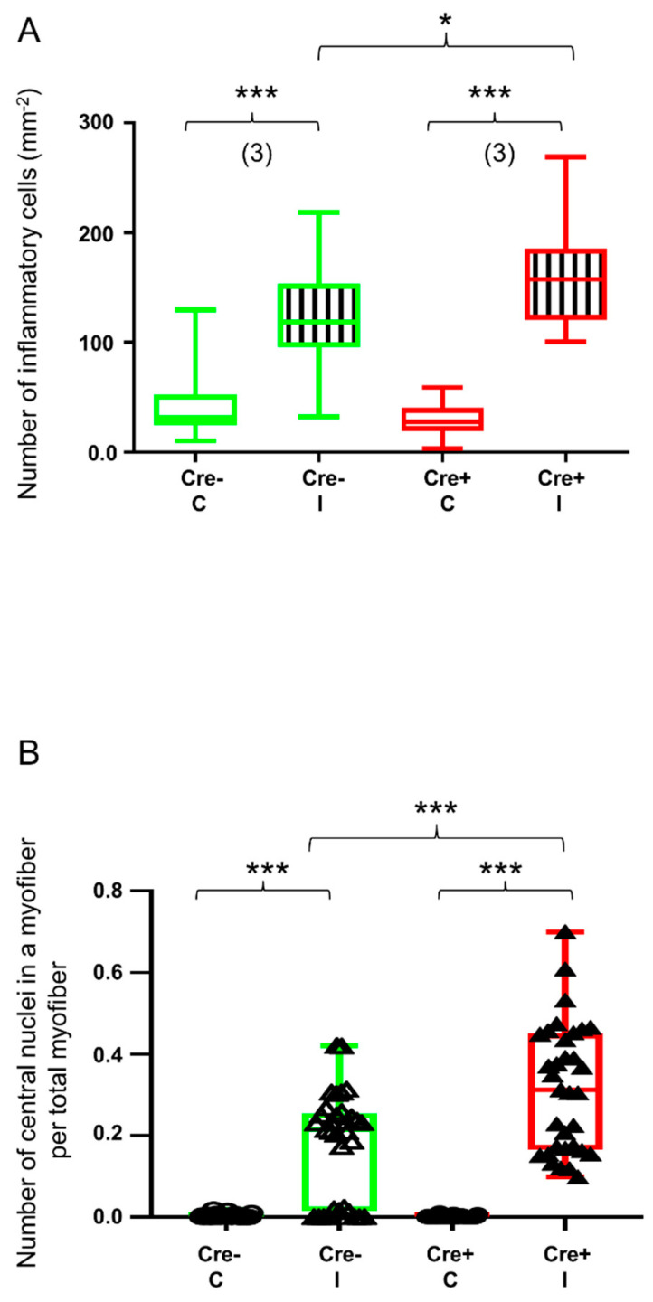
Figure 7.
Quantitative histological analysis shows delayed kinetics of regeneration in septin7 downregulated mice. Green color represents Cre− data and red color refers to Cre+ data. Numbers in brackets indicate the number of muscle samples. (A) Number of inflammatory cells per mm2. Inflammatory cells were counted on HE sections from Cre− and Cre+ mice 14 days after BaCl2 injection. Ten images were taken from one section, number of sections = 3 from all groups. (B) Number of centrally located nuclei in regenerating myofibers per total number of myofibers 14 days after injection. Asterisks indicate statistically significant difference (* p < 0.05, *** p < 0.001 Student’s t-test). Actual p value when p > 0.001 is (A) p = 0.024.

