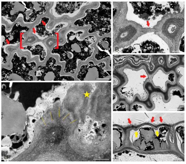
Figure 2.
Transmission electron micrographs of mature S. vulgare blades. (A) Tangential epidermal section, depicting the deeply wavy pattern of anticlinal walls. Note the local wall thickenings (some of which are pointed to by arrows) at the constricted wall sites (“necks”). (B) Higher magnification of the area defined by red brackets in (A). Two thickenings at opposite anticlinal walls are connected through the external periclinal wall by cellulose microfibril extension (arrow). (C) A tangentially sectioned epidermal cell, exhibiting a similar (compare with (A,B)) connection of opposite wall thickenings (arrow) through the external periclinal wall. (D) Tangentially sectioned epidermal cell at high magnification, depicting a local thickening at the junction of the anticlinal wall with the external periclinal one. Cellulose microfibrils in the thickening exhibit a radial orientation (labeled by lines). The asterisk notes a cellulose microfibril extension through the external periclinal wall, connecting with a neighboring thickening (in the right half of the micrograph). (E) Cross-section of an epidermal cell close to the junction of the external periclinal and anticlinal walls. Local thickening profiles can be observed (yellow arrows). The external periclinal wall exhibits protrusions (red arrows) in the areas between the thickenings. Scale bars (A): 5 μm, (B): 2 μm, (C): 5 μm, (D): 1 μm, (E): 5 μm.

