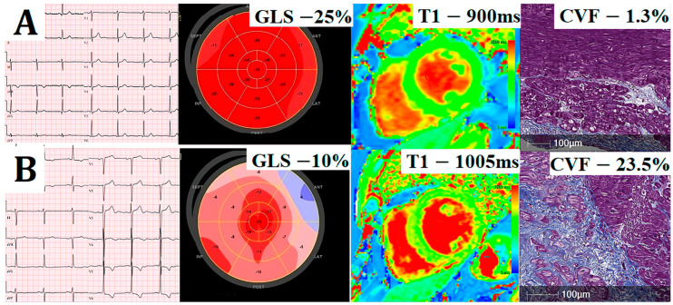
Figure 2.
Illustrative comparison of cardiovascular imaging and histology data of two exemplar patients: electrocardiography (Column 1), global longitudinal strain (GLS; Column 2), matching native T1 (Column 3), and collagen volume fraction (CVF) in myocardial biopsies stained with Masson’s trichrome (Column 4). Patient without ECG changes (A) has preserved GLS, low native T1, and low histological fibrosis (CVF of 1.3%), whereas patient with ECG strain (B) has significantly reduced GLS, high native T1, and extensive histological fibrosis (CVF 23.5%).

