Figure 9.
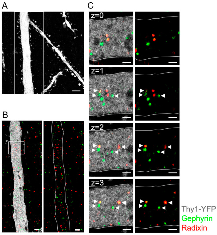
Expansion microscopy reveals gephyrin and radixin expression in dendrites of CA1 pyramidal neurons. (A) A single EYFP-positive dendrite of a CA1 pyramidal neuron in maximum z-stack projection is displayed after antibody staining against GFP (white). (B) Antibody staining against gephyrin (green) and radixin (red) in the boxed detail from (A) showed accumulation of gephyrin and radixin in the neuronal dendrite (GFP, white; single optical plane). The right panel displays gephyrin (green) and radixin (red) puncta in the cytoplasm of dendrite without the GFP signal. (C) Inset box from (B) at higher magnification. The primary dendrite showed localization of gephyrin and radixin in the cytoplasm (white arrowhead) in consecutive z planes. Note: co-localization of gephyrin and radixin in some clusters. Scale bar: (A): 2.5 µm; (B): 1 µm; (C): 500 nm. Z intervals = 0.5 μm post-expansion, 124 ± 7 nm pre-expansion.

