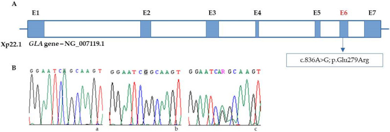
Figure 2.
(A) Localization of c.836A > G variant in the GLA gene (E = exon). (B) Chromatograms from Sanger sequencing of GLA in (a) control; Adenine (A) highlighted in grey, green single peak (b) hemizygous male, Guanine (G), highlighted in grey, black single peak; (c) heterozygous female, Guanine/Adenine, (R), double peak green/black.

