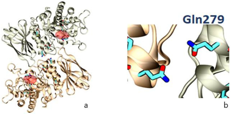
Figure 10.
Representation of the crystallographic structure of AGAL as a homodimer, with (a) the position of selected amino acids (Leu131, Asp 170, Gln279, and Met290) involved in FD (in blue) and the active site (in red); (b) localization of the amino acid, which is affected in this family (Gln279), as a result of the missense variant NM_000169.3:c.836A > G.

