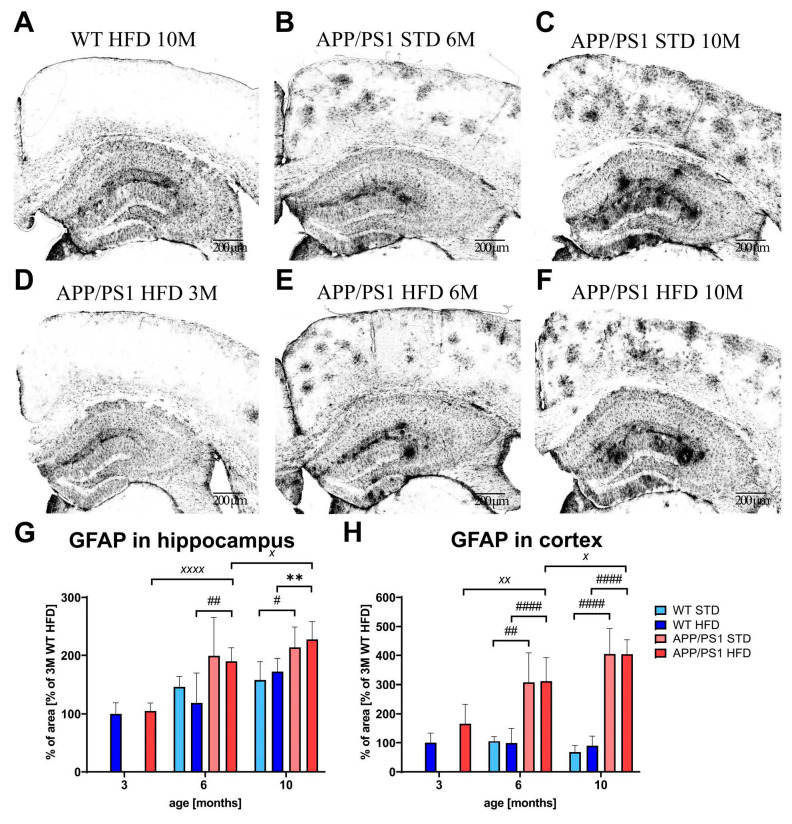
Figure 11.
Effect of HFD on astrocytosis in the hippocampi and cortices of the APP/PS1 mice. Representative photomicrographs of the brains of APP/PS1 mice fed either with STD at (B) 6 months or (C) 10 months or HFD at (D) 3 months, (E) 6 months and (F) 10 months of age and (A) the WT control at 10 months of age, immunohistochemically stained (A–F) for GFAP, and their quantification (G,H). Percentage of the stained area is expressed as a % of the 3-month-old WT mice on HFD to enable the comparison of multiple staining series. The data are presented as the means ± SD. Statistical analysis was made via one-way ANOVA with Bonferroni post hoc test (n = 5–8 mice per group). The significance of changes induced by age: X: p < 0.05, XX: p < 0.01, XXXX: p < 0.0001, by genotype: #: p < 0.05, ##: p < 0.01, ####: p < 0.0001, by diet: **: p < 0.01.

