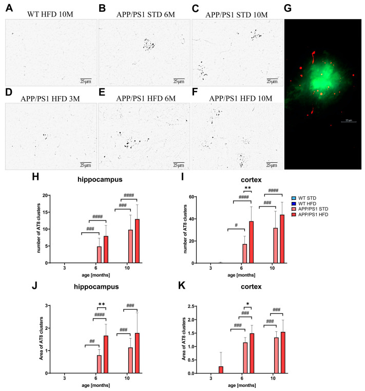
Figure 13.
Increased Tau phosphorylation around Aβ plaques in hippocampi and cortices of APP/PS1 mice. Representative photomicrographs of the APP/PS1 mice fed either with STD at (B) 6 months or (C) 10 months or HFD at (A) 3 months, (E) 6 months and (F) 10 months of age and (D) the WT control at 10 months of age, immunohistochemically stained (A–C) with AT8 antibody recognizing p-Tau at Ser202 and Thr205, and their quantification (H–K). (G) Representative figure of double staining of Aβ plaque (Thioflavin S) and p-Tau (AT8 antibody) in 10-month-old APP/PS1 mouse. Percentage of the stained area is expressed as a percentage of the 3-month-old WT mice on HFD to enable the comparison of multiple staining series. The data are presented as the means ± SD. Statistical analysis was made via one-way ANOVA with Bonferroni post hoc test (n = 5–8 mice per group). The significance of changes induced by diet: *: p < 0.05, **: p < 0.01, by genotype: #: p < 0.05, ##: p < 0.01, ###: p < 0.001, ####: p < 0.0001.

