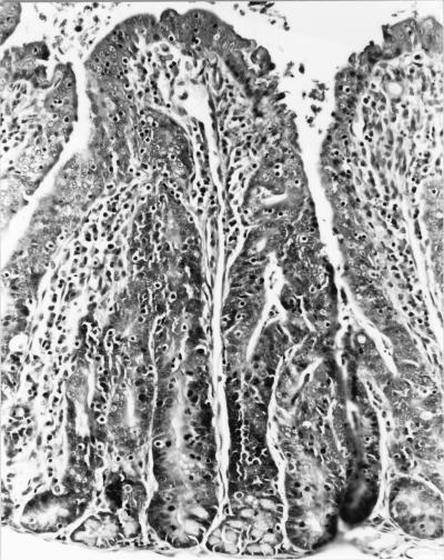
FIG. 1.
Photomicrograph of a section of small intestine from a rabbit with proliferative enterocolitis. The mucosa of the intestine has blunted villi, a moderate inflammatory cell infiltrate, and a decreased number of goblet cells. The crypt epithelial cells are markedly hyperplastic. Hematoxylin and eosin stain was used (magnification, ×40).

