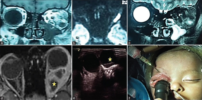
Figure 5.
(a and c) MRI orbit, coronal view, showing a well-circumscribed hypointense lesion on T1-weighted image and hyperintense lesion on T2-weighted image depicting a intraconal abscess in the inferolateral quadrant. (b) MRI orbit, axial view, diffusion-weighted image depicting a hyperintense signal, restricted diffusion along the lateral wall suggestive of an orbital abscess. (d) MRI orbits, axial view, T1-weighted, contrast-enhanced image depicts the classic multiseptate intraconal orbital abscess with rim enhancement. (e) Orbital mode B-scan ultrasonography. The star depicts a well-circumscribed hypoechoic lesion with a hyperechoic rim suggestive of a medial orbital abscess. (f) Peroperative clinical picture of a neonate depicting ultrasound-guided orbital abscess aspiration. MRI = magnetic resonance imaging

