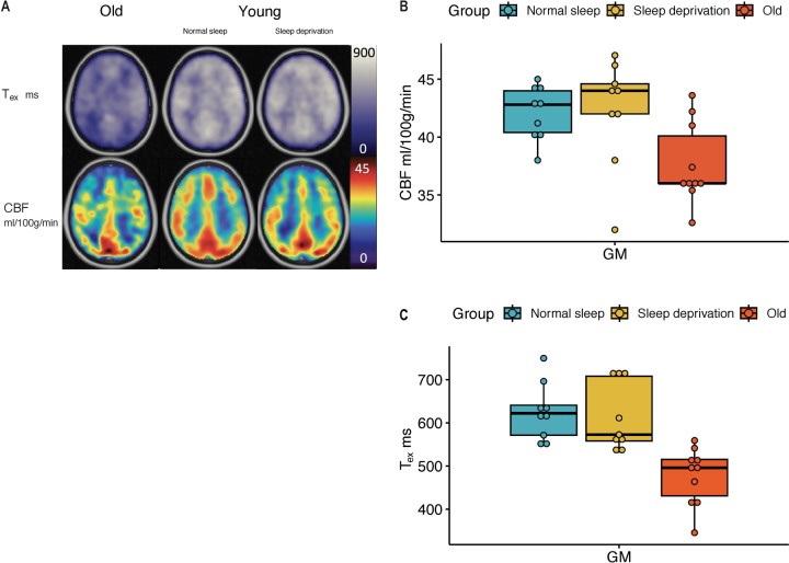
Figure 2:
A. ASL parameters, Whole-brain cortical CBF and Tex in older adults (first column), in young adults with normal sleep (middle column), and the same young adults with sleep deprivation (third column). CBF was not significantly different in young adults after sleep deprivation (yellow), when compared to those after a night of normal sleep (blue). Detailed paired comparison is shown in B. CBF was significantly different in older adults (red) compared to young adults (p=0.005). C. Tex was also not significantly different in young adults after sleep deprivation (yellow), when compared to those after a night of normal sleep (blue). Like CBF, Tex was significantly shorter in older adults (red) compared to young adults (p=0.0001).

