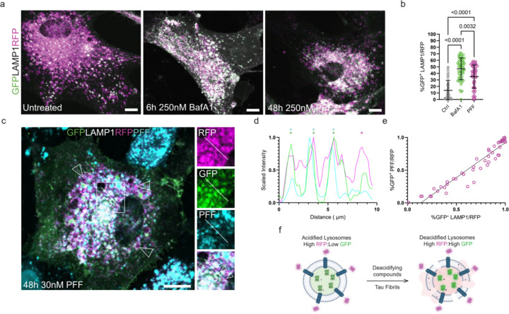
Figure 1: Tau fibrils permeabilize lysosomes.
Primary human astrocytes were stably transduced with a GFP-LAMP1-RFP tandem fusion protein to stoichiometrically monitor lysosomal pH. At rest, most lysosomes were RFP-positive. After treatment with Bafilomycin or PFF, the fraction of GFP-positive lysosomes increased dramatically (a). Quantification and 2way ANOVA of total cell GFP area normalized to total RFP signal prior to analysis (b) (n=52, 58, and 60 cells for untreated, BafA1, and PFF conditions, respectively, individual cells plotted with mean and standard deviation). The fibril seeding assay was performed in parallel using 30 nM fluorescently labeled PFF (c). A representative line scan from a cell with four neighboring lysosomes and only one containing labeled fibrils (RFP in magenta, GFP in green, and fibrils in cyan) (d). (e) Line plot revealing a positive linear correlation between cells that had GFP-positive lysosomes and GFP-positive PFF (R2=0.9215, n=50 cells, plotting individual Manders co-localization co-efficients). Schematic detailing the effect of deacidifying compounds and tau fibrils on the state of lysosomal pH (f). Scale bar: 20 µm.

