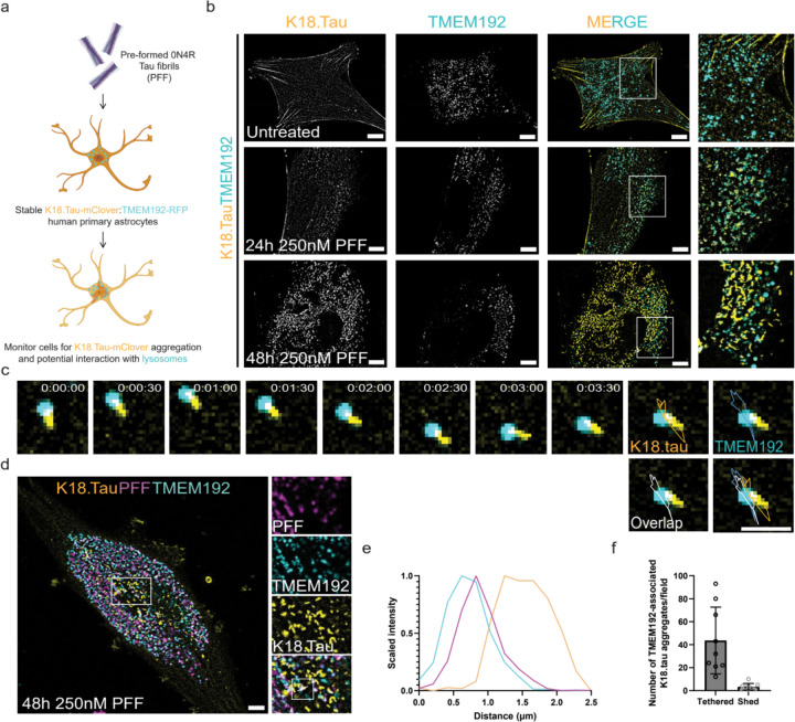
Figure 3: Soluble tau aggregation occurs in the proximity of lysosomes.
To model the tau fibril uptake mechanism by astrocytes in vitro, we treated astrocytes stably expressing K18.tau-mClover and TMEM192-RFP with PFF (a). These dual astrocytes were used in live imaging experiments to determine what impact PFF endocytosis had on the cytoplasmic pool of tau (b). At rest, cells had diffuse K18.tau expression that did not interact with lysosomes (top panel). After 24 hours of fibril treatment, K18.tau aggregates could be detected in close proximity to TMEM192-positive lysosomes (middle panel). The amount of K18 aggregation increased at 48 hours post-treatment with numerous lysosomes found associated with large K18 aggregates (lower panel). Representative trace of a lysosome-associated K18 aggregate during brief time-lapse imaging (c) (n=393). Representative cell from the PFF seeding assay repeated with fluorescently labeled PFF to verify lysosomal accumulation and association with K18 aggregates (d) (n=100). Fluorescence intensity plot of the line scan shown in (d), revealing the spatial distribution of PFF, lysosomes, and K18 aggregates (e). (f) Quantification of the number of lysosome-tethered versus shed tau aggregates (n=3 replicates, 3 random and independent fields of 20 cells per replicate, n=60 cells). Scale bars: 10 µm (b,d); 2 µm (c).

