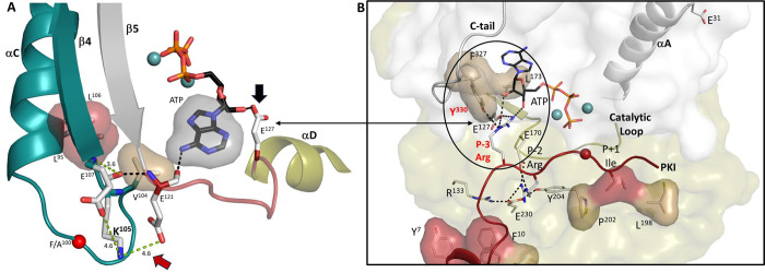
Figure 12. High-affinity binding of ATP and PKI converge at the αC-β4 loop.
(A). V104 in αC-β4 loop (in teal) is hydrophobically anchored to the adenine ring of ATP. The main chain carbonyl of K105 forms an H-bond to the backbone amide of E121, while its main chain carbonyl hydrogen bonds to ATP. The distance between K105 and E121 side chains is strengthened in the F100A mutant. Two spine residues, L95 and L106, in this loop are also shown. The linker that joins the N- and C-lobes (red) is flanked by E121 and E127. (B). Hydrophobic capping of the adenine ring of ATP is mediated mostly by N-lobe residues including V104 in the αC-β4 loop, as well as F327 in the C-terminal tail. In contrast, the P-3 to P+1 peptide is anchored to the catalytic machinery of the C-lobe. By binding to L173 in the C-lobe, the adenine ring completes the C-Spine, and thus fuses the adenine capping motif in the N-lobe with the extensive hydrophobic core architecture of the C-lobe. In the fully closed conformation, the side chain of Y330, also in the C-terminal tail, is anchored to the ribose ring of ATP. The only direct contact of the peptide/catalytic machinery with the N-lobe is mediated by the P-3 arginine which binds to the ribose ring of ATP and to E127 in the linker. In the fully closed conformation the P-3 arginine also binds to the side chain of Y330 in the C-terminal tail. Some of the mutations that disrupt the synergistic high-affinity binding of ATP and peptide/protein (E230Q, Y204A, F327A, L173A, and E31V) are highlighted. The hydrophobic residues in the amphipathic helix and P+1 inhibitor site of PKI are shown in red.

