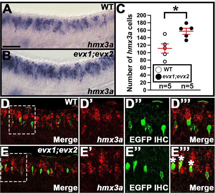
Figure 11. hmx3a Expression is Upregulated in a Subset of V0v Spinal Interneurons in evx1;evx2 Double Mutant Embryos.
(A, B, D-D”’, E-E”’) Lateral views of (A, D-D”’) wild-type and (B, E-E”’) evx1i232;i232;evx2sa140;sa140 double mutant embryos (labeled evx1;evx2) at 30 h. Rostral, left. Dorsal, up. (C) Number of cells expressing hmx3a in a precisely-defined spinal cord region adjacent to somites 6–10 at 30 h. Data are depicted as an individual value plot and n-values are indicated below. The wider red horizontal bar depicts the mean number of cells, and the red vertical bar depicts the S.E.M. (values are provided in Table 1). All counts are an average of five embryos. Statistically significant comparison is indicated with brackets and asterisks. * P <0.05. White circles indicate wild-type data and black circles indicate evx1;evx2 double mutant data. All data were first analyzed for normality using the Shapiro-Wilk test. Both data sets in C are normally distributed and so the F-test for equal variances was performed, followed by a type 2 Student’s t-test (for equal variances). P-values are provided in Table 1. (C) There is a statistically significant increase in the number of spinal interneurons expressing hmx3a in evx1;evx2 double mutant embryos. (D’, E’) in situ hybridization for hmx3a is shown in red. (D”, E”) Immunohistochemistry for Tg(evx1:EGFP)SU2, which exclusively labels V0v interneurons in the spinal cord (14), is shown in green. (D, D”’, E, E”’) Merged images. (D, E) Maximum intensity projection images. (D’-D”’, E’-E”’) High-magnification single confocal planes of the regions indicated by white dotted boxes in D and E. (E”’) A subset of ventral hmx3a-expressing cells in evx1;evx2 double mutant embryos co-expresses Tg(evx1:EGFP)SU2 (white asterisks in E”’), whereas there is no co-expression in the WT embryos (D”’). Scale bar: (A, B, D, E) 50 μm, (D’-D”’, E’-E”’) 35 μm.

