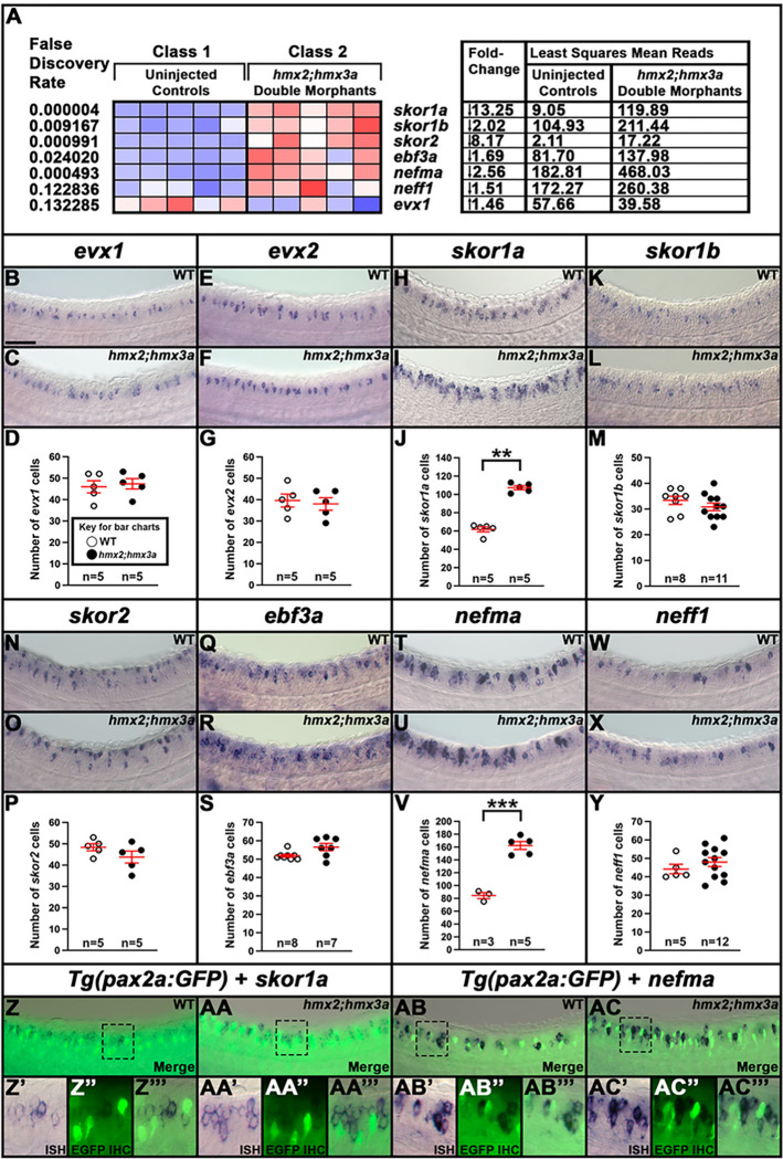
Figure 12. A Subset of V0v Spinal Interneuron Genes are Upregulated in hmx2;hmx3a4 Deletion Mutant Embryos.
(A) Heatmap analysis of gene-expression profiling of 27 h Tg(hmx CNEIII:cfos:Gal4:UAS:EGFP)SU41-expressing V1 and dI2 spinal cord interneurons. A two-class gene-specific analysis of differential expression was performed on different FAC-sorted populations of cells. Class 1: EGFP-positive cells from uninjected control embryos. Class 2: EGFP-positive cells from hmx2;hmx3a double knockdown (DKD) morpholino injected (morphant) embryos. Each column is a different biological replicate. Rows show relative expression levels for a subset of V0v candidate genes, shown as normalized data transformed to a mean of 0, with standard deviation of +1 (highly expressed, red) or −1 (weakly/not expressed, blue) sigma units. Adjusted P-values corrected for multiple testing (false discovery rate values) are shown on the left-hand side. Column 1 of right-hand table indicates fold-change reduction ( −) in uninjected controls compared to hmx2;hmx3a DKD morphant embryos. Columns 2 and 3 of right-hand table show least squares mean read counts for uninjected controls and hmx2;hmx3a DKD morphant embryos respectively. evx2 expression was not detected in either WT or morphant cells in this experiment. (B, C, E, F, H, I, K, L, N, O, Q, R, T, U, W, X, Z-AC”’) Lateral views of (B, E, H, K, N, Q, T, W, Z-Z”’, AB-AB”’) homozygous wild-type and (C, F, I, L, O, R, U, X, AA-AA”’, AC-AC”’) homozygous hmx2;hmx3aSU44;SU44 deletion mutant embryos at 27 h. Rostral, left. Dorsal, up. (D, G, J, M, P, S, V, Y) Number of cells expressing (D) evx1, (G) evx2, (J) skor1a, (M) skor1b, (P) skor2, (S) ebf3a, (V) nefma and (Y) neff1 in a precisely-defined spinal cord region adjacent to somites 6–10 at 27 h. Data are depicted as individual value plots with n-values provided below. For each plot, the wider red horizontal bar depicts the mean number of cells, and the red vertical bar depicts the S.E.M. (values are provided in Table 1). All counts are an average of at least three embryos. Statistically significant comparisons are indicated with brackets and asterisks. *** P <0.001. ** P <0.01. White circles indicate wild- type and black circles indicate data from homozygous hmx2; hmx3aSU44;SU44 mutants. All data were first analyzed for normality using Shapiro-Wilk test. Data sets in J and S are not normally distributed and Wilcoxon-Mann-Whitney tests were performed. Data sets in D, G, M, P, V and Y are normally distributed and so an F-test for equal variances was performed, followed by a type 2 Student’s t-test (for equal variances). P-values are provided in Table 1. (J, V) There is a statistically significant increase in the number of spinal interneurons expressing skor1a and nefma, but not (D, G, M, P, S, Y) evx1, evx2, skor1b, skor2, ebf3a, or neff1 in homozygous hmx2; hmx3aSU44;SU44 mutant embryos. in situ hybridization for (Z’, AA’) skor1a and (AB’, AC’) nefma genes is shown in dark blue. (Z”, AA”, AB”, AC”) Immunohistochemistry for Tg(pax2a:GFP), which specifically labels V1 interneurons in the spinal cord (6), is shown in green. (Z, Z”’, AA, AA”’, AB, AB”’, AC, AC”’) Merged images. (Z, AA, AB, AC) Maximum intensity projection images. (Z’-Z”’, AA’-AA”’, AB’-AB”’, AC’-AC”’) High-magnification single confocal planes of the regions indicated by black dotted boxes in Z, AA, AB, and AC. (AA”’, AC”’) The increased numbers of cells expressing (AA””) skor1a or (AC”’) nefma in (AA”’, AC”’) homozygous hmx2;hmx3aSU44;SU44 mutant embryos do not co-express Tg(pax2a:GFP), suggesting that the cells that have upregulated skor1a and nefma expression in these mutants are not V1 spinal interneurons. (B, C) evx1, (E, F) evx2 and (Q, R) ebf3a in situ hybridization experiments were performed with the molecular crowding reagent Dextran Sulfate. This was omitted for (H, I) skor1a, (K, L) skor1b, (N, O) skor2, (T, U) nefma and (W, X) neff1 in situ hybridization experiments. Scale bar: (B, C, E, F, H, I, K, L, N, O, Q, R, T, U, W, X, Z, AA, AB, AC) 50 μm, (Z’-Z”’, AA’-AA”’, AB’-AB”’, AC’-AC”’) 20 μm.

