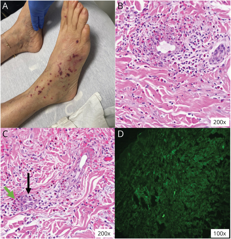
Figure 2. Histopathologic Features.

(A) Palpable nonblanching lesions suggestive of cutaneous vasculitis. (B and C) Skin biopsy showing eosinophilic infiltration (green arrow) around vessels (black arrow). (D) On direct immunofluorescence, skin vessels stained positive for antibodies against IgG, IgM, C3, and fibrinogen; intercellular basement membrane and superficial dermis stained negative for antibodies.
