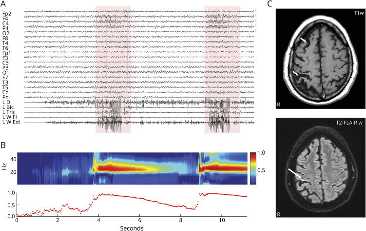
Figure 1. EEG-Polygraphic Recording, CMC Analysis, and Brain MRI.
(A) Two reflex seizures over the right central leads associated with muscular bursts (boxes). (B) Sudden increase of C4/left wrist flexor muscles CMC2 during voluntary movement to seizure shift. (C) Brain MRI shows (top) right hemisphere atrophy and (bottom) signal hyperintensity in the right postcentral gyrus (arrows). CMC = corticomuscular coherence.

