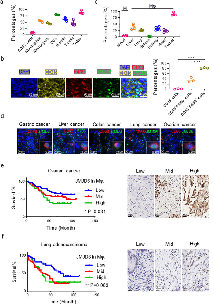
Fig. 1. JMJD6 is upregulated in TAMs, and correlates with the cancer prognosis.
a JMJD6 expression in tissue macrophages (monocytes in blood) was detected by flow cytometry, n = 4. b Representative immunofluorescence images of tumor sections from mice subcutaneously implanted with LLC cells, labeled by CD45 (yellow), F4/80 (red), JMJD6 (green) and DAPI (blue). Scale bars are marked in individual images. c JMJD6 expression in tumor cells (CD45-), tumor-infiltrating neutrophils (CD45+ CD11b+ Ly6G− Ly6C+), monocytes (CD45+ CD11b+ Ly6G− Ly6C+), DCs (CD45+ CD11b+ F4/80− CD11c+ MHC-II+), T cells (CD45+ CD3+), B cells (CD45+ CD19+), and TAMs (CD45+ CD11b+ F4/80+) was detected by flow cytometry, n = 4. d Representative immunofluorescence images of tumor tissues from clinical samples. As marked in images, sections were labeled with CD68 (red), JMJD6 (green), DAPI (blue). Scale bars are marked in individual images. Survival curves of ovarian cancer patients (e) and lung adenocarcinoma patients (f) grouped by JMJD6 expression in stromal cells (left panel), analyzed by Logrank test for trend. Immunohistochemistry images were representative regions of JMJD6 expression in the stromal cells (right panel). Data are represented as mean ± SD. *P < 0.05, **P < 0.01.

