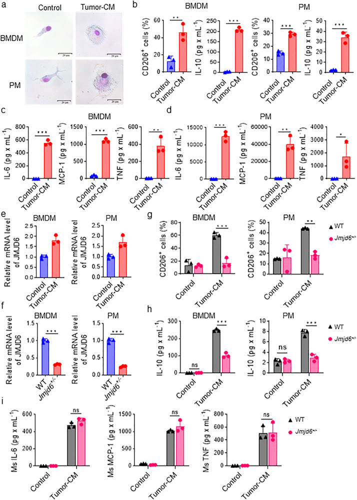
Fig. 2. JMJD6 is upregulated in macrophages stimulated by tumor-conditioned medium and promotes macrophage activation.
a Representative images after Giemsa staining of BMDM and PM treated by normal medium or tumor-CM. b CD206 and IL-10 expression on BMDMs and PMs treated by normal medium or tumor-CM were analyzed by flow cytometry and cytometric beads array (CBA), respectively. CBA was conducted to measure expression level of IL-6, MCP-1, IL-10 and TNF in the supernatant of the TC-Mφ from BMDMs (c) and PMs (d). e mRNA expression of JMJD6 was detected by qPCR in PMs and BMDMs stimulated by tumor-CM or not. f mRNA expression of JMJD6 was detected in TC-Mφ from BMDMs and PMs of WT or Jmjd6+/− mice. g Flow analysis reveals the expression of M2 biomarkers CD206 in BMDMs and PMs stimulated by tumor-CM or not. h The secretion level of IL-10 in BMDMs and PMs stimulated by tumor-CM or not was detected by CBA assays. i BMDMs from WT or Jmjd6+/− mice were cultured in Tumor-CM for 48 h, and the secretion level of IL-6, MCP-1, and TNF was detected by CBA. Data represent mean ± SD (n = 3). ns: no significant difference, *P < 0.05; **P < 0.01; ***P < 0.001.

