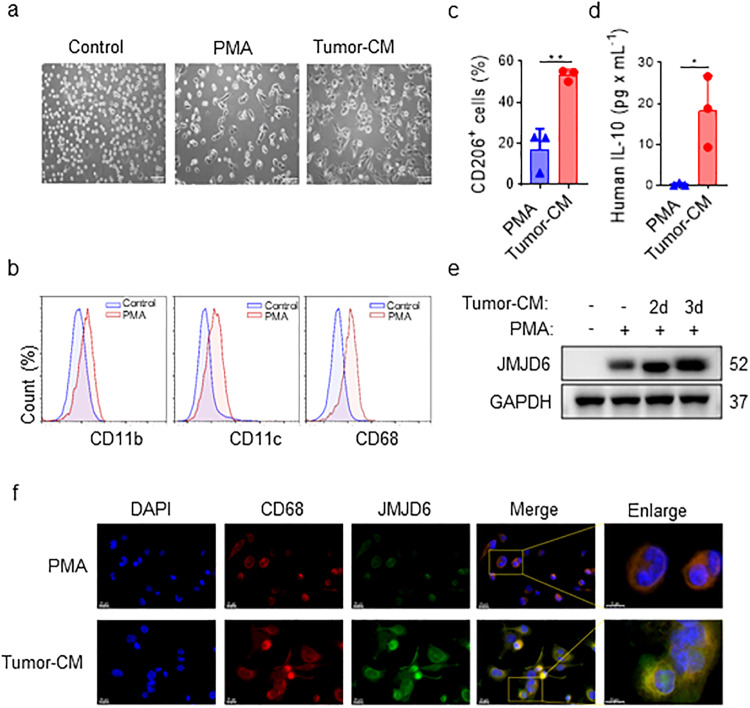
Fig. 4. Tumor-CM activates THP-1 cells and upregulates JMJD6 expression.
a Morphology of THP-1 cells cultured in normal medium, with or without PMA and tumor-CM (H460) for 48 h. Scale bar, 100 μm. b The changes of macrophage related markers on the surface of THP-1 cells were detected by flow cytometry after PMA induction. Representative results. c CD206 expression of TAM surfaces were detected by flow cytometry after tumor-CM induction. d The secretion of IL-10 in the TAM culture supernatant detected by CBA assays. e THP-1 cells were treated with PMA alone or tumor-CM for 2 or 3 days, followed by JMJD6 protein level detection. f Representative images of immunofluorescence staining of CD68 (red), JMJD6 (green) and DAPI (blue) in THP-1 cells treated with PMA alone or tumor-CM for 2 days, the scale bars 10 μm and 20 μm (right panel) as indicated in figures. Data are shown as mean ± SD (n = 3). *P < 0.05, **P < 0.01.

