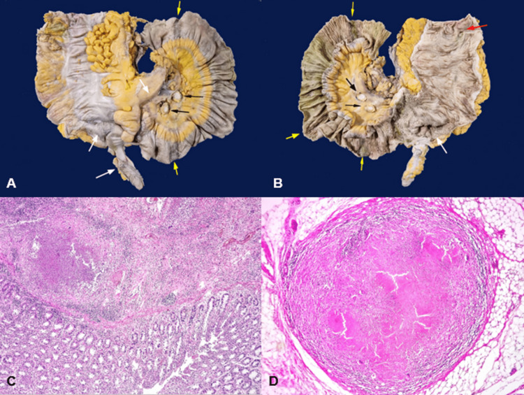
Figure 2. Pathological anatomy.
(A) Serosal surface of the terminal ileum, cecal appendix, cecum, and ascending colon. Yellow arrows indicate areas perpendicular to the longitudinal axis of the intestine, dark brown in color, with multiple light brown pinpoint lesions (miliary pattern) in the terminal ileum. White arrows show multiple light brown pinpoint lesions (miliary pattern) in the cecal appendix, cecum, and mesentery. Black arrows indicate the cut surface of two mesenteric lymph nodes with caseous necrosis.(B) Mucosal surface of the terminal ileum, ileocecal valve, cecum, and ascending colon. Yellow arrows show ulcers perpendicular to the longitudinal axis of the intestine in the terminal ileum, associated with the serosal surface changes mentioned in A. The largest ulcer measures 2.0 cm in width and involves the entire intestinal circumference, while the smallest measures 1.0 × 0.3 cm. The white arrow shows an ulcer affecting the ileocecal valve and part of the cecum, measuring 7.5 × 7.0 cm. The red arrow shows a 2.5 cm × 1.0 cm ulcer in the ascending colon. (C) (hematoxylin and eosin (H&E) 40×) Colonic mucosa with abundant acute and chronic inflammatory infiltrate, and in the submucosa, a suppurative granuloma is identified, consisting of central necrosis, acute inflammatory infiltrate, epithelioid cells, some multinucleated giant cells, and lymphocytes at the periphery. (D) (H&E 40×) Tuberculoid granuloma. Well-circumscribed lesion showing central caseous necrosis, epithelioid cells, intercalated multinucleated giant cells, and lymphocytes at the periphery.

