Fig. 3.
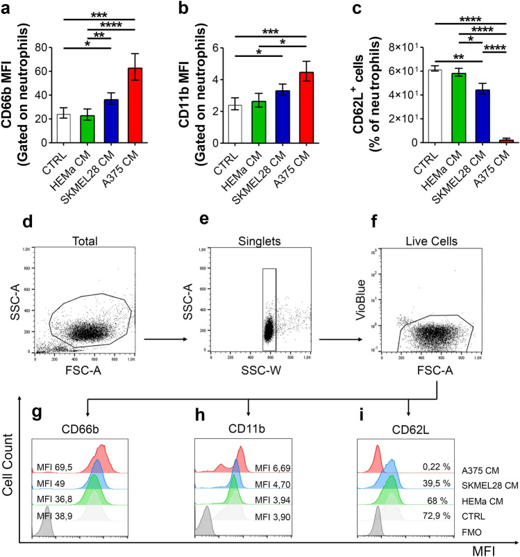
Melanoma-derived CM induced PMN activation. a–c PMNs were stimulated with melanoma CM (SKMEL28 CM, A375 CM), HEMa CM or control medium for 90 min at 37 °C, stained for neutrophil activation markers CD66b (a), CD11b (b) and CD62L (c) and subjected to cytofluorimetric analysis. The results were expressed as mean fluorescence intensity (MFI) or percentage of positive cells gated on neutrophils (mean ± SEM of seven independent experiments); one-way ANOVA and Dunn’s multiple comparison test; *p < 0.05; **p < 0.01; ***p < 0.005; ****p < 0.001. d–f Representative flow cytometric panels were gated on live single cells and show forward (FSC) and side scatter (SSC) of EasySep-purified untouched neutrophils (d, e). Since Vioblue-positive cells included both dead cells and CCR3+ cells (eosinophils), both cells were excluded based on a negative gate (f). g–i Representative histograms illustrating MFI and cell count for CD66b (g), CD11b (h) and CD62L (i) for one out of seven experiments. MFI mean fluorescence intensity, FMO fluorescence minus one

