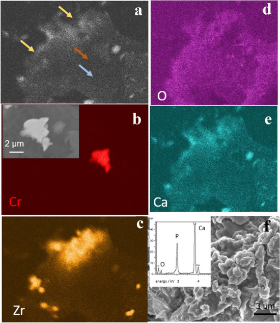
Fig. 8.

Morphology and composition of wear particles isolated from periprosthetic tissue by alkaline protocol. a SEM BSE image showing particles differing in composition (heavy elements appear darker than bright ones), as pointed out by arrows. b–f EDS compositional maps of the SEM image in Co, Zr, O, and Ca. Arrows of the same colour note the presence of particles in the SEM image in (a). SEM BSE image of the metal particle is shown in the inset in (b). f SEM SE image and EDS spectrum of calcium phosphate particles
