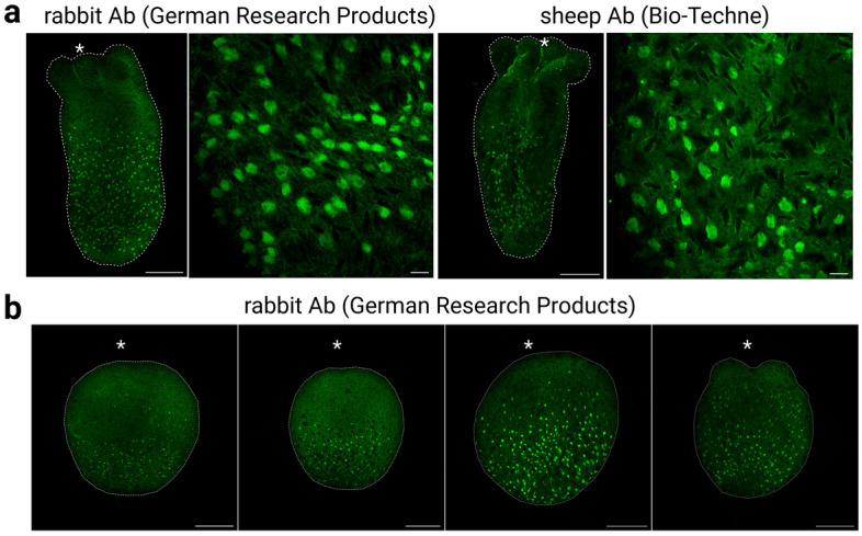
Figure 5.

Morphology of NvEAAT1-positive cells. (a) EAAT1-expressing cells stained with German Research Products (rabbit) and Bio-Techne (sheep) anti-EAAT1 antibodies are broadly detected throughout the aboral region of the body column during the juvenile polyp stage (9dpf). Cells have flat morphology and lack processes. (b) Expression of NvEAAT1 at different developmental stages revealed by immunostaining with German Research Products anti-EAAT1 antibody. EAAT1-expressing cells became visible during larval stages. Side views of (from left to right): late gastrula stage (3dpf), early planula stage (4dpf), planula stage (5dpf), tentacle bud stage (6dpf). Asterisks indicate oral positions. Large and small scale bars are 100 μm and 10 μm, respectively.
