Figure 6.
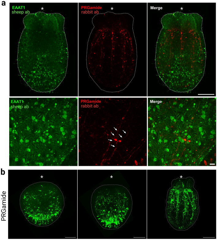
Non-overlapping expression of PRGamide and EAAT1 protein in the N. vectensis nervous system. (a) Immunofluorescence co-staining using anti-EAAT1 antibody (Bio-Techne) and anti-PRGamide antibody during the primary polyp stage (7dpf). EAAT1 staining and PRGamide staining do not overlap. EAAT1-positive cells lack processes, unlike PRGamide-positive neurons, which extend neurites (arrows). (b) PRGamide-expressing neurons detected at different developmental stages. Side views of (from left to right): early planula stage (4dpf), planula stage (5dpf), juvenile polyp stage (7dpf). Asterisks indicate oral positions. Large and small scale bars are 100 μm and 10 μm, respectively.

