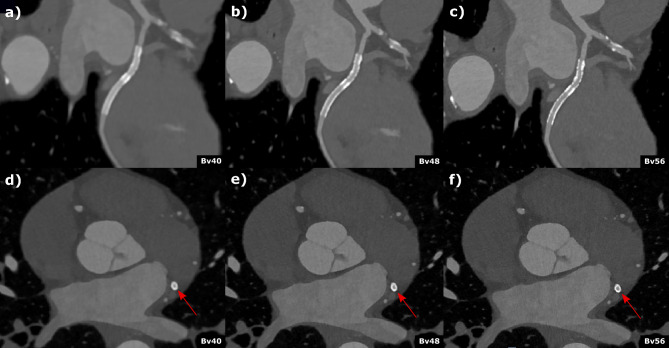
Figure 4.
Coronary computed tomography angiography of a 62-year-old male patient with known coronary artery disease and stenting of the circumflex artery. Curved (a–c)) multiplanar reformations and axial (d–f)) reconstructed with different kernel strength (a and d) Bv40, (b and c) Bv48, (c and f) Bv56 depict the stent (2.5 mm diameter) in the circumflex artery. Stent lumen was best visible in the Br56 kernel reconstruction (c and f) and an in-stent restenosis could be reliably excluded. Scan was performed with 144 × 0.4 mm dual source, multi spectral high-pitch-flash-mode (3.2). Effective radiation dose was 1.07 mSv.

