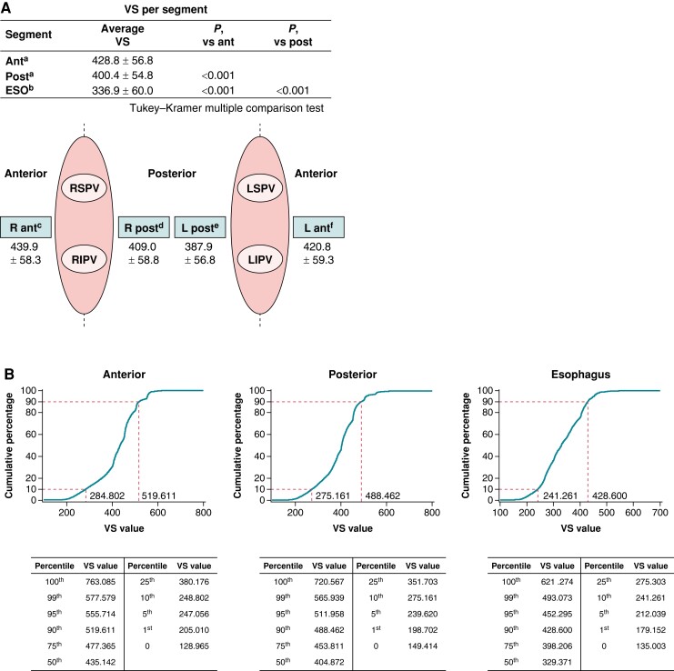
Figure 2.
Details of the ablation procedure (A) and cumulative distribution of VS values in the anterior, posterior, and oesophagus regions (B). Ant, anterior; ESO, oesophagus; LIPV, left inferior pulmonary vein; LSPV, left superior pulmonary vein; post, posterior; RIPV, right inferior pulmonary vein; RSPV, right superior pulmonary vein; VS, VISITAG SURPOINT. an = 965, bn = 651, cn = 954, dn = 955, en = 952, and fn = 960.

