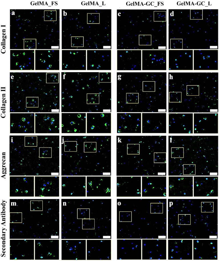
FIG. 4.
Extracellular matrix in free-swelling (FS) and uniaxially loaded (L) constructs after 28 days of culture. Immunofluorescence staining for (a)–(d) collagen I, (e)–(h) collagen II, (i)–(l) aggrecan, and (m)–(p) secondary antibody of statically cultured and loaded GelMA (15 %, w/v) and GelMA-GC (GelMA 15% and GC 1%, w/v) hydrogels cross-linked by Ru/SPS for 5 min at 405 nm LED light. Immunoreactive regions for collagen I, collagen II, and aggrecan appear green. Secondary antibody as a negative control appear green. Nuclei were counterstained with DAPI (blue). Scale bar: 200 μm. White boxes on main images are shown at higher magnification directly below the main image.

