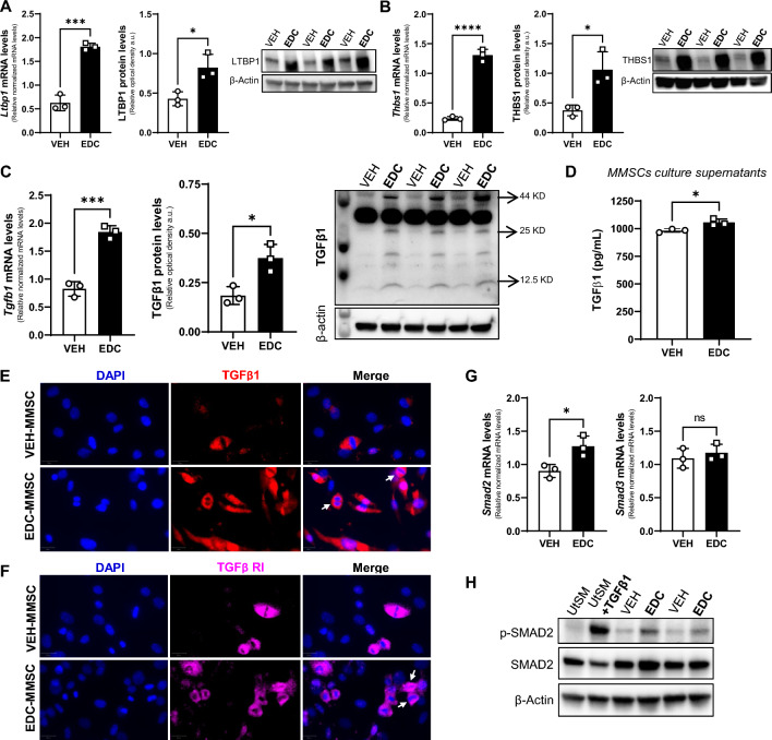
Fig. 1.
The TGFβ1 pathway is overactivated in MMSCs isolated from rats neonatally exposed to EDC. Real-time PCR analysis of mRNAs, protein levels and representative gels of A LTBP1, B THBS1, and C TGFβ1 (arrows indicate band for: Pro-TGFβ1 at 44 kDa, Dimer of mature TGFβ1 at 25 kDa, and Monomer of mature TGFβ1 at 12.5 kDa) in VEH- and EDC-MMSCs isolated from 5-month-old rats. D TGFβ1 levels in culture supernatants collected from VEH- and EDC-MMSCs cultures. Immunofluorescence images of E TGFβ1 and F TGFβ RI in VEH- and EDC-MMSCs. Scale bar 20 µm. The white arrows indicate cells in mitotic stages expressing TGFβ1 or TGFβ RI. F mRNAs levels of Smad2 and Smad3 in VEH- and EDC-MMSCs isolated from 5-month-old rats. G Representative gel of p-Smad2 and Smad2 in VEH- and EDC-MMSCs. mRNA data were normalized by the amount of 18S and protein levels by the amount of β-actin. Data are shown as mean ± S.E.M. from triplicate data. ns not significant. *p < 0.05, ***p < 0.001, ****p < 0.0001, Student’s t test. EDC endocrine-disrupting chemical, MMCSs myometrial stem cells, THBS1 thrombospondin 1, LTBP1 latent TGFβ binding protein 1, TGFβ1 transforming growth factor beta 1, TGFβ RI transforming growth factor beta receptor 1, UtSM human uterine smooth muscle cell line. TGFβ1 treatment: 10 ng/ml for 1 h (positive control)

