Fig. 2.
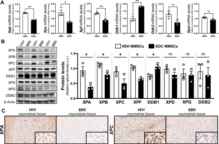
Characterization of nucleotide excision repair (NER) pathway in VEH- and EDC-MMSCs. A mRNA levels of Xpb, Xpc, Xpf, Ddb1, Ddb2, and Xpd in VEH- and EDC-MMSCs isolated from 5-month-old rats. 18S was used to normalize the expression data. B Representative gel and protein levels of XPA, XPB, XPC, XPF, DDB1, XPD, XPG, and DDB2 in VEH- and EDC-MMSCs isolated from 5-month-old rats. Data were normalized by the amount of β-actin protein levels. C IHC images (×20 magnification, insets are at ×40 magnification) of XPA and XPC in myometrial tissues from 5-month-old Eker rats treated neonatally with VEH or EDC. Scale bar 200 µm. Data are shown as mean ± S.E.M. from triplicate data. *p < 0.05, **p < 0.01, Student’s t test. EDC endocrine-disrupting chemical, MMSCs myometrial stem cells, XP xeroderma pigmentosum, DDB1/2 DNA damage-binding protein 1/2

