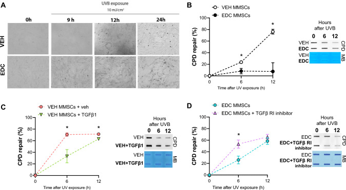
Fig. 3.
Effect of early life EDC exposure and TGFβ1 on CPD repair. A Bright-field images of VEH- and EDC-MMSCs before (0 h) and after UVB exposure (9, 12, and 24 h; 10 mJ/cm2). Magnification ×20. B Quantification of percentage (%) of CPD repair and a representative image of DNA slot blot in VEH- and EDC-MMSCs isolated from 5-month-old rats at 0, 6 and 12 h post-UVB (10 mJ/cm2). C Quantification of percentage (%) of CPD repair and a representative image of DNA slot blot in VEH-MMSC treated with vehicle (4 mM HCl + 0.1% BSA) or TGFβ1 (10 ng/ml) for 48 h and then collected at 0, 6 and 12 h post-UVB (10 mJ/cm2). D Quantification of percentage (%) of CPD repair and a representative image of DNA slot blot in EDC-MMSC treated with vehicle (< 0.1% DMSO) or TGFβ1 receptor inhibitor (2 µM) for 24 h and then collected at 0, 6, and 12 h post-UVB (10 mJ/cm2). Methylene blue (MB) was used as the loading control. Data are shown as mean ± S.E.M. from triplicate data. *p < 0.05, Student’s t test

