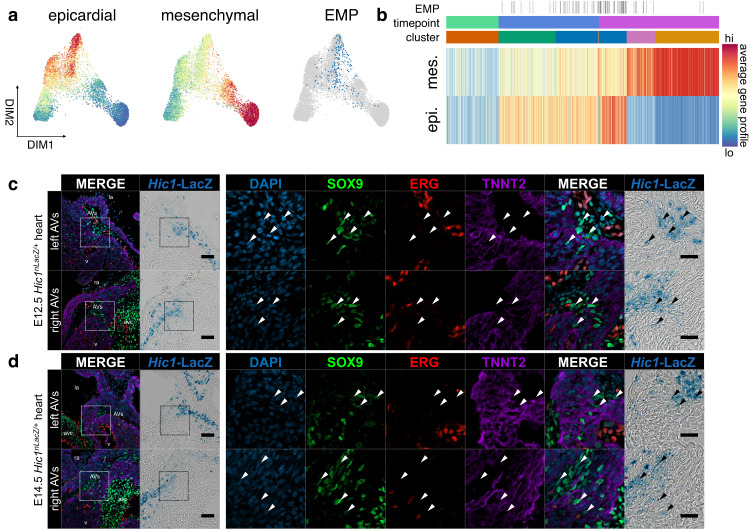
Fig. 6. Epithelial-mesenchymal plasticity during EpiMT.
a Force-directed layouts and (b) a heatmap of 4633 epicardial cells displaying average epicardial and mesenchymal gene profiles. Overlap in gene profile expression suggests emergence of EMP during EpiMT. Immunostaining of Hic1nLacZ/+ AV canals for SOX9, ERG, and TNNT2 at (c) E12.5 (n = 9) and (d) E14.5 (n = 6) shows co-expression of mesenchymal SOX9 and AV EPDC Hic1nLacZ within AV sulcus, suggesting transition to mesenchyme (arrowheads). Scale bars: 100 µm, 25 µm in ROIs. AV atrioventricular, AVs AV sulcus, epi epicardium, mes mesenchyme. See also Supplementary Fig. 6 and Supplementary Data 2 and 4.

