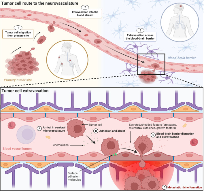
Fig. 2.
The origin of brain metastases at the blood–brain barrier. Tumor cell route to the neurovasculature. 1 Tumor cells at the primary site accumulate attributes that enable the manipulation of the surrounding tissue, allowing for cell migration and tissue invasion. 2 Tumor cells reach blood vessels at the primary site and intravasate across the vessel wall into the blood stream. 3 Circulating tumor cells reach the neurovasculature, arrest on the endothelium and extravasate across the blood–brain barrier. Tumor cell extravasation. A Tumor cells arrive in the neurovasculature, where chemokines are able to facilitate attraction to the brain endothelium. B Arrest on the brain endothelium is supported by vessel dynamics, as well as surface adhesion molecules located on both the endothelial and tumor cell membrane. C Various secreted and shedded factors released by the tumor cell disrupt the blood–brain barrier and facilitate extravasation across the brain endothelium. D After extravasation, disseminated tumor cells remain in the perivascular microenvironment where they continue to release factors that support the development of a metastatic niche. Created with BioRender.com

