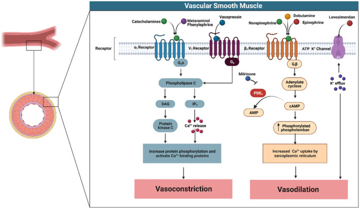
Figure 2. Intracellular signaling within vascular smooth muscle following the activation of α1‐adrenergic, vasopressin‐1, β2‐adrenergic, and ATP‐sensitive potassium channel receptors.

Stimulation of the α1‐adrenergic or vasopressin‐1 receptors causes activation of the Gq (G‐protein) subunit, resulting in downstream stimulation of the phospholipase C signaling pathway. Phospholipase C in turn activates inositol 1, 4, 5‐triphosphate (IP3) leading to Ca2+ release from the sarcoplasmic reticulum (SR), resulting in vasoconstriction. Conversely, β2‐receptor stimulation activates the inhibitory G‐protein (Gi) subunit causing vasodilatation through increased cAMP (cyclic adenosine monophosphase) activation and resulting phospholamban‐mediated Ca2+ uptake into the SR. Similarly, activation of the ATP‐sensitive potassium channel causes potassium influx that hyperpolarizes voltage‐dependent Ca2+ channels, reducing intracellular Ca2+ and vasomotor tone. AMP indicates adenosine monophosphate; DAG, diacylglycerol; PDE3, phosphodiesterase 3; and PLB, phospholamban.
