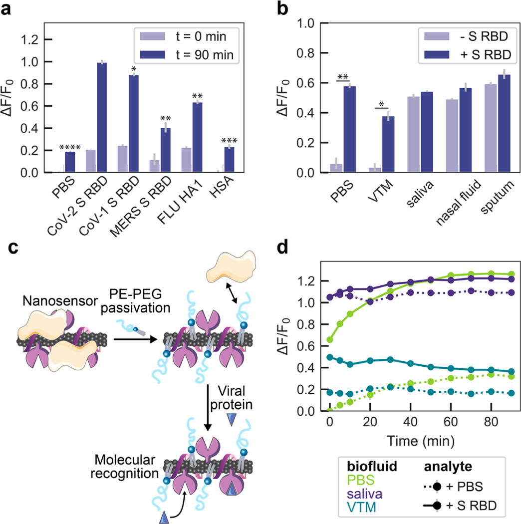
Figure 3.
ACE2-SWCNT nanosensor selectivity and sensitivity in biofluid environments. (a) Normalized change in fluorescence (ΔF/F0) of the 1130 nm SWCNT emission peak for the ACE2-SWCNT nanosensor 0 and 90 min after exposure to 10 mg/L of viral protein panel: SARS-CoV-2 spike receptor-binding domain (S RBD), SARS-CoV-1 S RBD, MERS S RBD, and FLU hemagglutinin subunit (HA1). ****P = 0.0006 (PBS), ***P = 0.0014 (HSA), **P = 0.0065 (FLU) and 0.0076 (MERS), and *P = 0.0503 (SARS-CoV-1) in independent two-sample t tests, for each analyte ΔF/F0 response at t = 90 min in comparison to SARS-CoV-2 S RBD. (b) ACE2-SWCNT nanosensor response 90 min after exposure to 1 μM S RBD in the presence of 1% relevant biofluids: viral transport medium (VTM), saliva, nasal fluid, and sputum (treated with sputasol). **P = 0.0065 (PBS) and *P = 0.0161 (VTM) in independent two-sample t tests, for ΔF/F0 response in biofluids compared before vs after S RBD addition. (c) Schematic depiction of nanosensor biofouling with proteins present in relevant biofluids, mitigated upon passivation with phosphatidylethanolamine phospholipid with a 5000 Da PEG chain (PE–PEG). (d) Response of PE–PEG passivated nanosensor to 500 nM S RBD in the presence of PBS, 10% VTM, or 1% saliva. Surface passivation with a hydrophilic polymer improved the nanosensor response that was otherwise greatly attenuated, as shown in (b). All fluorescence measurements were obtained with 721 nm laser excitation. Gray bars represent the standard error between experimental replicates (N = 3).

