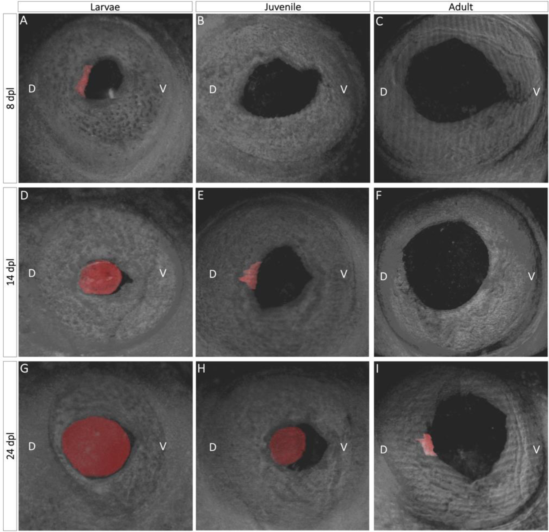
Fig. 2:

Three-dimensional view of lens regeneration. B- scans were used to reconstruct a three-dimensional image of the newt eye. To aid in visualization, the regenerating lens was pseudo-colored with red color. Three-dimensional images permit detailed observations of lens growth and morphogenesis. When it first appears, the lens vesicle is located in the mid-dorsal region of the iris and has an asymmetrical shape (A, E, I). As the lens develops and more lens epithelial cells differentiate into lens fibers, the regenerating lens assumes a spherical shape (D, G, H).
