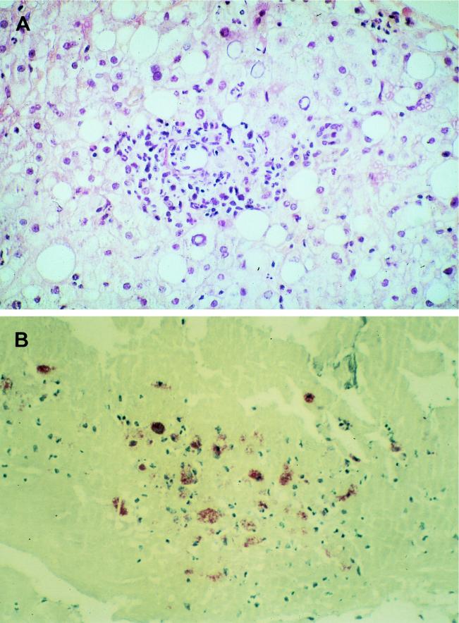
FIG. 2.
(A) Liver in acute Q fever. A liver biopsy specimen was stained with hematoxylin-phloxin-saffron. One granuloma is seen within the fatty liver parenchyma. The lesions consist of inflammatory infiltrates made of epithelioid cells, polymorphonuclear leukocytes, and histiocytes. Magnification, ×400. (B) Immunoperoxidase staining of a cardiac valve biopsy specimen showing fibrous valvular tissue comprising inflammatory infiltrates made of histiocytes. Magnification, ×40.

