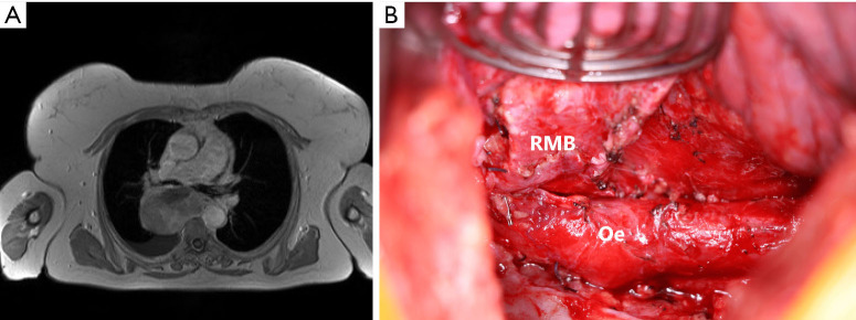
Figure 3.
Preoperative imaging and intraoperative situs in a patient with leiomyosarcoma of the middle mediastinum. (A) MRI shows a 90 mm leiomyosarcoma of the middle mediastinum. (B) Intraoperative picture after leiomyosarcoma resection. The RMB and Oe were not infiltrated by the tumor. MRI, magnetic resonance imaging; RMB, right main bronchus; Oe, oesophagus.

