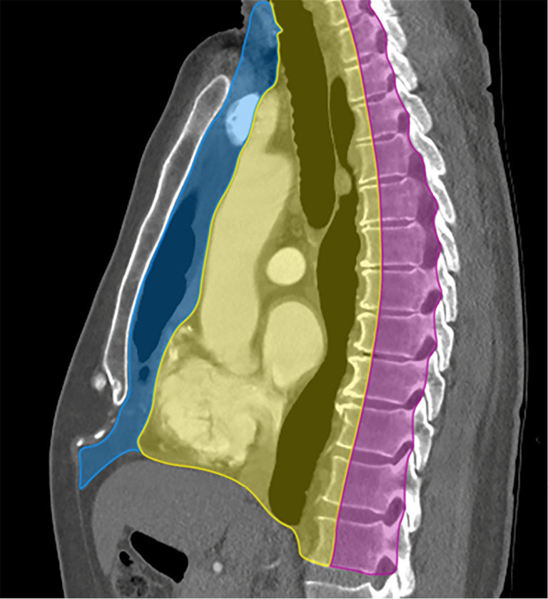
Figure 1.

ITMIG classification of mediastinal compartments bound superiorly by the thoracic inlet and inferiorly by the diaphragm. Sagittal CT with intravenous contrast shows the prevascular compartment (blue), bound anteriorly by the sternum and posteriorly by the anterior aspect of the parietal pericardium, and contains the thymus, fat, lymph nodes, and the left brachiocephalic vein. The visceral compartment (yellow) is bound anteriorly by the posterior boundaries of the prevascular compartment, and posteriorly by a vertical line 1 cm posterior to the anterior margin of each thoracic vertebral body, and contains the trachea, esophagus, lymph nodes, and vascular structures including the heart, thoracic aorta, superior vena cava, intrapericardial pulmonary arteries, and the thoracic duct. The paravertebral compartment (pink) is bound anteriorly by the posterior boundaries of the visceral compartment, and posterolaterally by a vertical line against the posterior margin of the chest wall at the lateral margin of the transverse process of the thoracic spine. ITMIG, International Thymic Malignancy Interest Group; CT, computed tomography.
