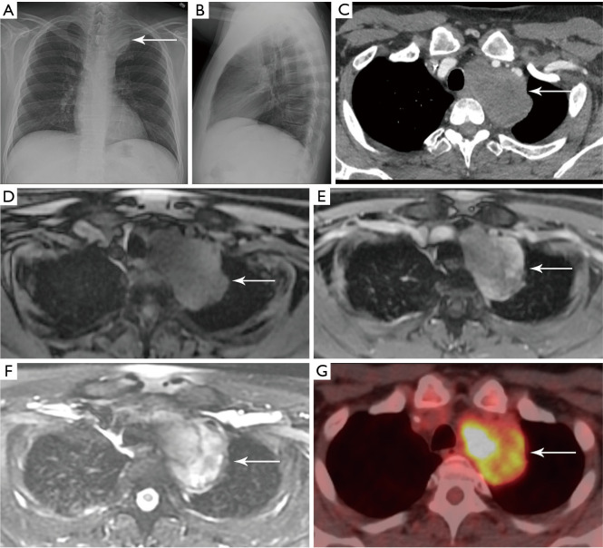
Figure 11.
Schwannoma. (A,B) PA and lateral chest radiographs show a left mediastinal mass (arrow) in the upper thorax. (C) Axial contrast enhanced CT shows the mass (arrow) is solid with heterogeneous attenuation. (D) Axial T1-weighted MRI shows the mass (arrow) is iso-intense to muscle. (E) Axial T1-weighted post-contrast MRI shows the mass (arrow) enhances heterogeneously. (F) Axial T2-weighted MRI shows the mass (arrow) is heterogeneously hyperintense with cystic areas. (G) Fused PET/CT shows the mass (arrow) is FDG avid. Biopsy confirmed schwannoma. PA, posterior anterior; CT, computed tomography; MRI, magnetic resonance imaging; PET, positron emission tomography; FDG, fluoro-2-deoxy-D-glucose.

