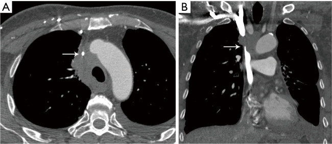
Figure 12.
SVC obstruction. (A) Axial contrast enhanced CT shows mediastinal adenopathy due to lung cancer narrowing the SVC (arrow) with multiple collaterals in the right anterior chest wall. (B) Coronal contrast enhanced CT shows the SVC is patent below the level of obstruction due to the adenopathy (arrow). SVC, superior vena cava; CT, computed tomography.

