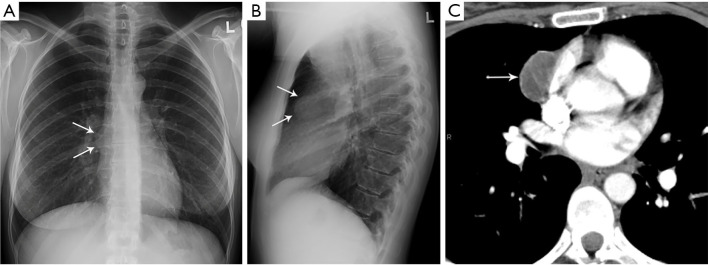
Figure 2.
Pericardial cyst. (A,B) Frontal and lateral chest radiograph shows subtle contour abnormality (arrows) with loss of the silhouette of part of the right mediastinal border termed the “silhouette sign”. (C) CT shows low attenuation consistent with pericardial cyst (arrow). CT, computed tomography.

