Figure 7.

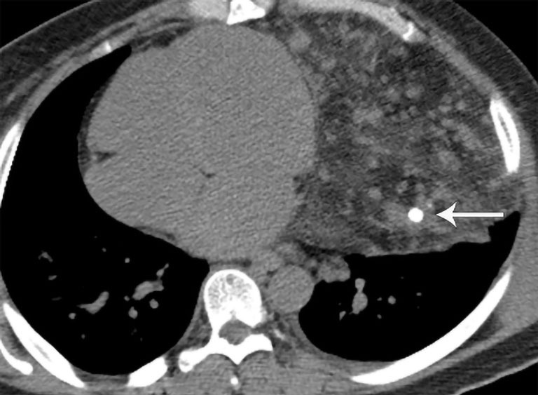
Hemangioma. CT shows large left mediastinal mass with focus of calcification (arrow) consistent with phlebolith interspersed with soft tissue and fat. CT, computed tomography.

Hemangioma. CT shows large left mediastinal mass with focus of calcification (arrow) consistent with phlebolith interspersed with soft tissue and fat. CT, computed tomography.