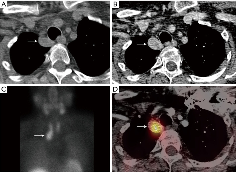
Figure 9.
Ectopic parathyroid adenoma. (A,B) Pre- and post-contrast enhanced CT show right paratracheal solid lesion (arrow) with heterogeneous enhancement. The pre-contrast CT is useful to differentiate high attenuation thyroid tissue from low attenuation parathyroid tissue. (C,D) Technetium-99m sestamibi parathyroid scintigraphy and SPECT/CT show the right parathyroid adenoma is sestamibi avid (arrow). CT, computed tomography; SPECT, single photon emission computed tomography.

