FIGURE 4.

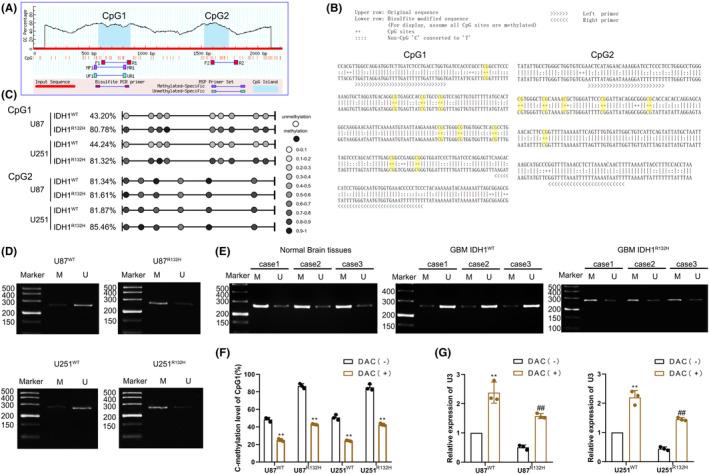
Promoter hypermethylation inhibits U3 expression in IDH1WT GBM. (A) U3 promoter CpG islands were analyzed using the MethPrimer program. (B) Schematic representation of CpG1 and CpG2 bisulfite sequencing and primers. (C) Methylation status of CpG1 and CpG2 at the U3 promoter was detected in IDH1WT and IDH1R132H GBM cells via bisulfite genomic sequencing. (D) Methylation status of CpG1 at the U3 promoter was detected via methylation‐specific PCR in IDH1WT and IDH1R132H GBM cells (M, methylated; U, unmethylated). (E) Methylation status of CpG1 at the U3 promoter was detected via methylation‐specific PCR in normal brain tissues, IDH1WT GBM tissues, and IDH1R132H GBM tissues (M, methylated; U, unmethylated). (F) Change in methylation level of CpG1 in IDH1WT and IDH1R132H GBM cells after treatment with 5‐aza‐2′‐deoxycytidine (DAC). **p < 0.01 versus DAC(−) group. (G) U3 expression was detected in IDH1WT and IDH1R132H GBM cells after treatment with DAC. **p < 0.01 versus IDH1WT DAC(−) group, ## p < 0.01 versus IDH1R132H DAC(−) group. Data are presented as the mean ± SD of three independent experiments per group, unless otherwise specified. The data were statistically analyzed via the Student's t‐test.
