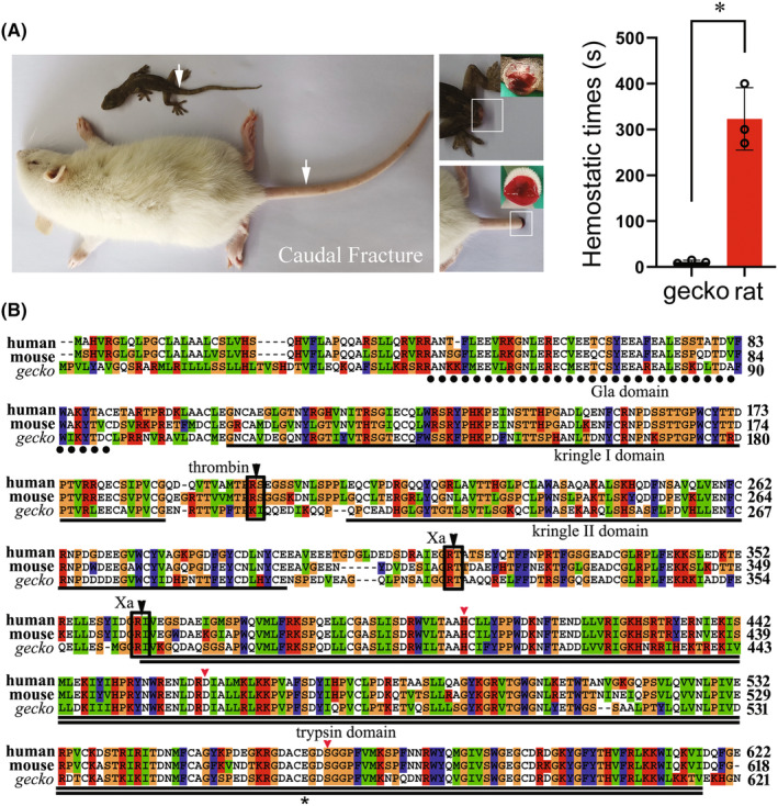
FIGURE 1.

Gross observation of gecko wounding hemostasis and analysis of amino acid sequence of gecko prothrombin. (A) Comparison of gecko and rat natural hemostatic time following tail amputation. The tail of gecko was amputated above 6th, while the rat at 9‐11th caudal vertebra. (B) Multiple alignment of amino acid sequences of gecko prothrombin with those of human and mouse. Each residue in the alignment is assigned a color if the amino acid profile of the alignment at that position meets some minimum criteria specific for the residue type. Gaps introduced into sequences to optimize alignment are represented by dashes. The Gla domain, kringle I domain, kringle II domain, and trypsin domain are indicated by dot line, line, or double line, respectively. The potential cleavage sites by coagulation factor Xa and thrombin are boxed and indicated by the black arrowhead. The conserved catalytic residues, His91, Asp147, and Ser251 (numbering from the amino terminus of the light chain), are indicated by the red arrowhead. The Glu248 for gecko and Glu251 for human, around which the negative electrostatic potentials are analyzed, are indicated by the asterisks. Prothrombin sequences of gecko (XP_015262498), human (AAC63054), and mouse (NP_034298) are obtained from GenBank.
