Figure 1.

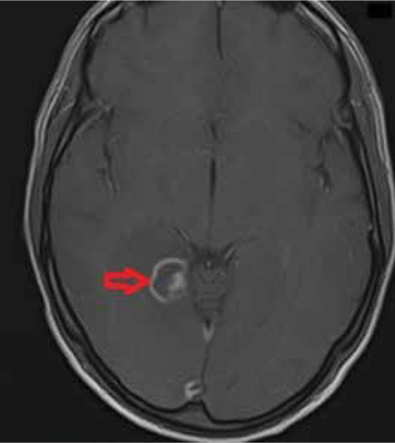
T1-weighted axial post-contrast image shows a lesion with ring enhancement and an additional peripheral nodular enhancement within the lesion; this is referred to as an eccentric target sign typical in neurotoxoplasmosis

T1-weighted axial post-contrast image shows a lesion with ring enhancement and an additional peripheral nodular enhancement within the lesion; this is referred to as an eccentric target sign typical in neurotoxoplasmosis