
Figure 2.
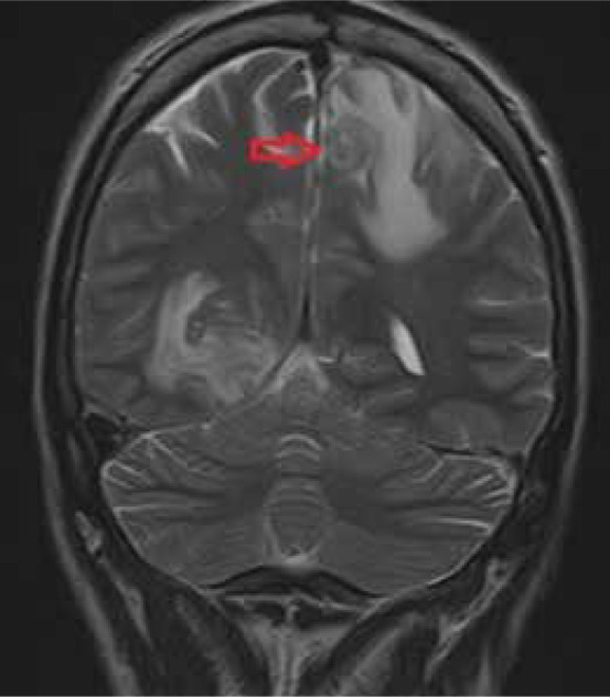
Coronal T2-weighted MRI of the brain shows a concentric alternating zone of hypo-/hyper /isointense signal, surrounded by perilesional oedema, which, with the accompanying T1 post-contrast images, are typical features of neurotoxoplasmosis
Official websites use .gov
A
.gov website belongs to an official
government organization in the United States.
Secure .gov websites use HTTPS
A lock (
) or https:// means you've safely
connected to the .gov website. Share sensitive
information only on official, secure websites.
Coronal T2-weighted MRI of the brain shows a concentric alternating zone of hypo-/hyper /isointense signal, surrounded by perilesional oedema, which, with the accompanying T1 post-contrast images, are typical features of neurotoxoplasmosis