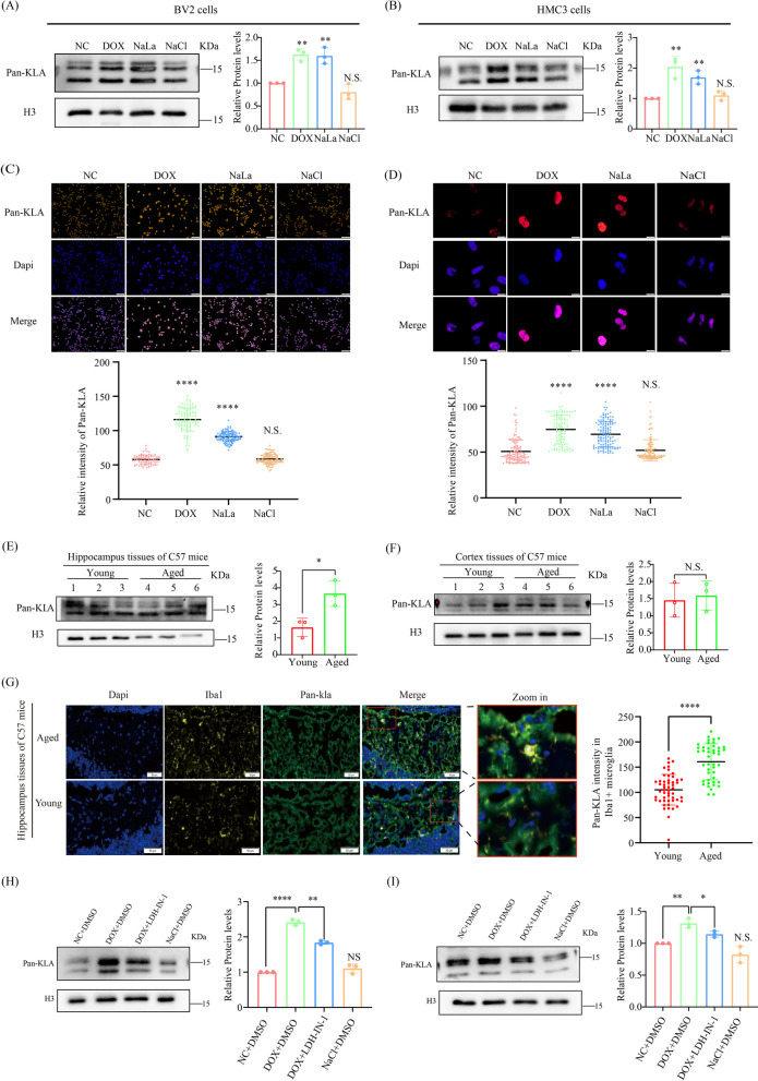
Fig. 2.
Histone Pan-Kla levels is increased in premature senescent microglia and hippocampus tissues of naturally aged mice. A, B Measurement of histone Pan-Kla levels in the indicated microglia groups by immunoblotting assay. C, D Detection of histone Pan-Kla levels in the indicated microglia groups by immunofluorescence. E Measurement of Pan-Kla levels in hippocampus tissues from young and naturally aged mice (n = 3 mice per group). F Multiplexed immunohistochemistry measurement of Pan-Kla levels in hippocampus tissues from young and naturally aged mice (n = 3 mice per group), scale bar, 50 μm. G Immunoblotting analyzes for Pan-Kla levels in the indicated microglia groups. Each bar represents the mean ± s.d. for triplicate experiments, *p < 0.05, **p < 0.01, ***p < 0.001, N.S.: no significance

